
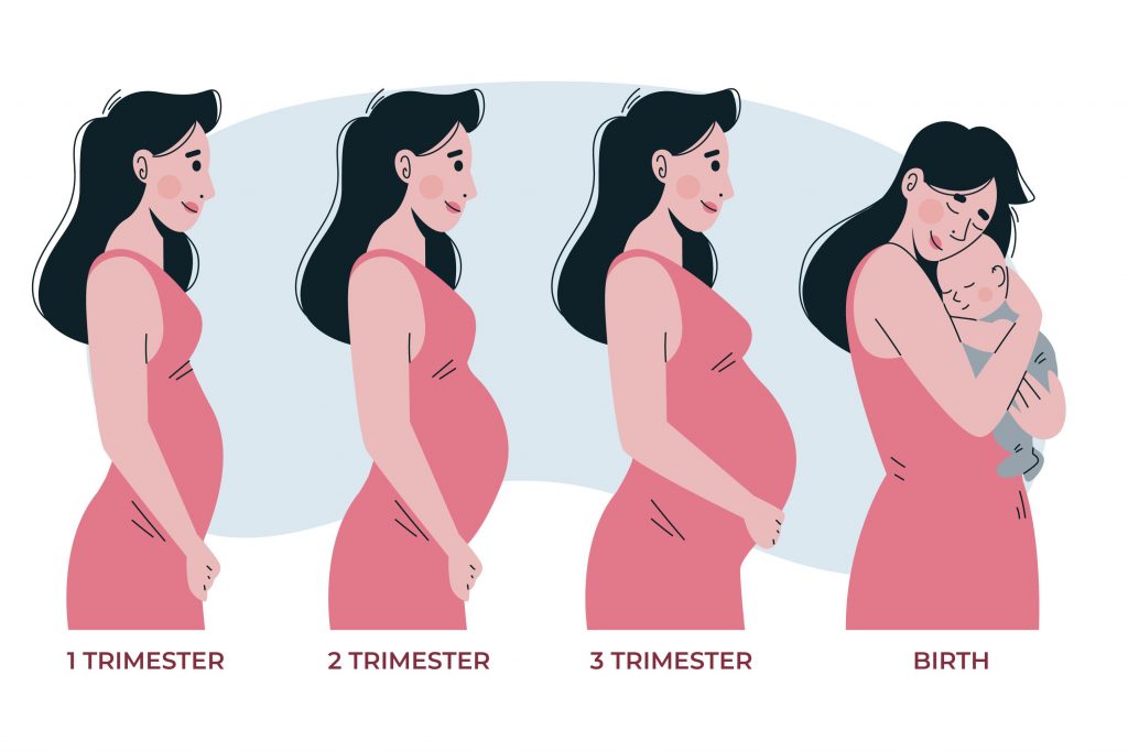
Pregnancy and Pregnancy Tracking
Please consult your gynecologist and obstetrician.
Pregnancy follow-up with gynecological examination before myrtle;
Evaluation of the uterus and ovaries
Investigation for the presence of infection
Performing a smear test (cervical cancer test)
Fasting blood sugar, whole blood test, kidney and liver function tests
Previous drug use and history of persistent disease should be investigated.
Folic acid should be started 3 months before conception.
Your period is late
Pregnancy test is positive in blood or urine. Consult your doctor.
At First Control;
It is examined with ultrasound (most often vaginal ultrasound). In the early weeks of pregnancy and pregnancy follow-up, better results are obtained with vaginal ultrasound compared to abdominal ultrasound. Vaginal ultrasound has no harm to pregnancy. It does not cause miscarriages.
If the Beta-Hcg value is below 1500, the increase should be monitored every 2 days. If the value does not increase by the required amount, conditions such as ectopic pregnancy (pregnancy developing outside the uterus), miscarriage should be considered.
If the Beta-Hcg value is over 1500, vaginal usg, if it is over 6500, the gestational sac can now be seen with abdominal usg. In the 6th week of ultrasonography, the baby’s heartbeat can be monitored.
In the first control, blood group, complete blood count, liver and kidney function tests, urine tests, hepatitis B, hepatitis C, AIDS, tests related to infections that are dangerous to be passed during pregnancy are requested.
Second Control (11-14 Weeks)
During this period, diseases related to chromosomal disorders such as Down Syndrome (Tr21), Tr18, Tr 13 should be screened.
Down Syndrome is a disease that occurs when the 21st chromosome should come from the mother and father, but 2 from the mother. The incidence increases if the maternal age is over 35. This syndrome is a condition in which mental retardation and some internal organ and heart anomalies are seen together.
With ultrasound, the baby’s head-butt distance and nape skin thickness are measured. Nasal bone (nasal bone) measurement is made. Afterwards, it is combined with free Beta-Hcg and PAPP-A values in the mother’s blood to determine the probability of having Down syndrome in the baby, and pregnancy and pregnancy follow-up are important here. Amniocentesis or chorionic villus biopsy is recommended for high-risk results. Amnicentesis is the process of taking a sample of the fluid in the baby by entering with a needle from the mother’s womb. Chorionic villus biopsy is the process of genetic examination by taking a piece of the placenta (partner) of the baby. Amniocentesis has a 1/100 – 1/1000 probability of causing abortion. It is 99% diagnostic.
Third Control (16-18 Weeks)
In ultrasound, the fetus is generally evaluated for anomaly. Whether there is an opening between the vertebrae in the back region, stomach, arms and legs, the whole body is evaluated in general. In the triple test performed during this period, the hormones called Beta-Hcg, AFP and Estriol (E3) are checked from the mother’s blood. Again, the aim is to determine the risk of chromosomal anomaly in the baby. If the risk is high as a result of the test, amniocentesis is recommended. The rate of detection of Down Syndrome of the test is 60%.
With the addition of inhibin-A hormone in the triple test, the chance of success of the test was increased. It is known as the quadruple test because of the fact that four hormones are checked in the mother’s blood. Down Syndrome detection rate is 80%.
With the triple and quadruple test, it can also be understood whether there is a gap between the vertebrae in the baby’s back area.
According to the results of amniocentesis, babies with Down Syndrome can be terminated with the request of the family, by means of pregnancy and pregnancy follow-up.
In recent years, free fetal DNA tests have become available in amniocentesis and chorionic villus biopsy methods due to the risk for mother and baby. In this test, cell particles belonging to the baby are examined in the mother’s blood. It can be done in any period from the 10th week of pregnancy. The blood taken from the arm is examined in the mother. Results are obtained within 7-10 days. The accuracy of the test is 99% for trisomy 21 and 18. The accuracy rate for trisomy 13 is 80%. However, this test is not a definitive diagnostic test. It is a screening test. should be confirmed and a definitive diagnosis should be made. As Mersin, we are here for you to lead a healthy life.
Fourth Control (20-24 Weeks)
With USG, the baby’s internal organs, brain, heart and arms and legs are examined in detail. Fetal anomalies are investigated.
Fifth Control (24-28 Weeks)
In pregnancy and pregnancy follow-up, it is investigated whether there is anemia and hidden infections in the urine in pregnant women, and treatment is given if necessary. Between these weeks, a sugar load test with 75 g must be done. With this test, it is determined whether the mother has gestational diabetes. There is no harm in the test. Diet or insulin therapy is started depending on the situation in pregnant women with high gestational diabetes. If the treatment is not done, the baby may be overweight and have difficult births due to this, and the development of permanent diabetes in the baby and the mother may be encountered. If the values are high, the diagnosis is made.
Indirect Coombs test; If the mother is RH- and the father is Rh+, RH incompatibility can be seen. If the blood test called indirect Coombs test is negative, Anti-D Ig is administered to the mother at 28 weeks. In addition, if the baby is Rh + after birth, Anti D Ig is repeated to the mother.
From the 32nd week, follow-up is done every two weeks. In USG, amniotic fluid, placenta and active movements of the baby are examined. NST and fetal Doppler USG are used to detect risky situations.
NST (Nonstress test): It shows the course of the baby’s heartbeat and uterine contractions. It is checked whether there is a decrease in heart rate with contractions or baby movements. It takes 20-30 minutes. This procedure does not have a negative effect on the mother or the baby.
Color Doppler USG: It measures the blood flow to the baby. Blood flow disorders in the baby’s umbilical cord and brain vessels are detected. Thus, the deterioration of the baby’s condition is detected and follow-up or delivery is planned according to the situation. Pregnancy and pregnancy follow-up is very important in this process.